Want to learn about internal turkey anatomy? Read on to discover the structure and internal turkey anatomy…
It is important to assess the internal turkey anatomy if you are involved in poultry farming. This is required because you need to see if the bird has some diseases in its body. By checking internal turkey anatomy and being familiar with its original structure you will be able to check the organ systems for any changes occurring due to disease.
If you know what the normal external and internal anatomy of the turkey should look like it is very easy to discover upon examination if you have some sort of diseased bird in your poultry farm.
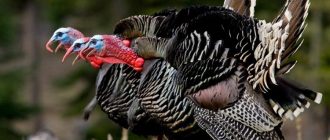
As far as the external anatomy is concerned there is a lot of plumage on the body of the turkey which follows feathered tracts rather than being randomly present there.
These are normally clear and clean at the root attached to the body. While the skin is thin and semitransparent and you can see the veins, fat deposits along with the muscles of the turkey. You should be able to identify the dark muscular areas and the yellow fatty areas.
Internal Turkey Anatomy
Remove the skin on one of the birds and expose the internal organs completely. Each organ in a healthy bird should be evaluated properly so that you know what a healthy organ is.
The first thing you will be able to see is the muscle, sternal bursa and the keel bone. The healthy breast muscles are grayish white in color and the pointed end of the keel should be white and its edge should be very straight. The sternal bursa is the saclike structure which is located at the sternum and is white in color. It should be filled with clear fluid.
When you observe the muscles in the leg they will be comparatively darker and grayish white in color. The sciatic nerve which is located between the muscles of the leg is shiny white and has crossed striations.
When you observe the chest and abdominal cavity you will have to remove the sternum and breast muscles. The heart of the turkey is a triangular shape and the base faces upwards towards the head. It is surrounded by the pericardial sac which is clear. The heart should be grayish white in color and the pointed end at the bottom should have a fatty deposit encircling it.
If you slice the heart open it should have a monochromatic color and very clear membranes separating the heart chambers. Notice that the left ventricle is thicker than the right ventricle. The liver lobes almost completely encircle the heart.
In the turkey you will find that the liver has very sharp edges and is the largest organ in the internal turkey anatomy. The color depends on the diet. Baby poults normally have a yellowish liver because of the absorption of the yolk whereas adult turkeys have a tan to yellowish colored liver if the diet is rich in fat. However a balanced diet could result in a reddish brown colored liver.
The liver lobe has the turkeys gallbladder attached to it and this is greenish black in color because of the bile in it. The spleen is normally 1 inch long and is oval in shape and has a reddish purple color. Healthy lungs are attached close to the ribs and have a pinkish red color.